
Inverted V Deformity
The inverted V deformity is a recognised post-rhinoplasty complication that can also occur following nasal trauma. It refers to the visible appearance of the caudal (lower) edges of the nasal bones through the overlying skin, creating a shadow pattern on the nasal bridge that resembles an inverted letter V when viewed from the front. It is a sign of middle vault collapse and can affect both the appearance and the function of the nose.
All surgery carries risks — read the full rhinoplasty risks page →
What Causes an Inverted V Deformity?
The normal nasal dorsum transitions smoothly from the nasal bones in the upper third to the upper lateral cartilages in the middle third. The upper lateral cartilages are connected to both the nasal bones above and the nasal septum medially. When a dorsal hump is removed, these connections are disrupted — the roof of the nose is opened and the natural supporting relationship between these structures is lost.
If the middle vault is not adequately reconstructed at the time of dorsal reduction — using spreader grafts or spreader flaps — the upper lateral cartilages lose their lateral support and collapse inward toward the septum. As this happens, the lower edges of the nasal bones become more prominent, and the characteristic inverted V shadow appears on the bridge.
Beyond the cosmetic effect, middle vault collapse narrows the internal nasal valve, which is the most important single determinant of nasal airflow. The inverted V deformity is therefore frequently associated with nasal obstruction.
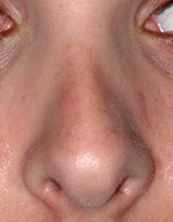
Inverted V deformity — visible nasal bone edges from middle vault collapse
Prevention
The inverted V deformity is largely preventable. When a dorsal hump reduction is performed, spreader grafts should routinely be placed at the same time to reconstruct the middle vault and maintain the support of the upper lateral cartilages. Patients at highest risk — those with short nasal bones, thin skin, and long upper lateral cartilages — are particularly important to protect with spreader grafts at the time of dorsal reduction.
If you are planning rhinoplasty that involves dorsal reduction, ask your surgeon specifically whether spreader grafts are planned and why or why not.
Treatment
The primary treatment for an established inverted V deformity is spreader grafts — placed through a revision rhinoplasty to reopen the middle vault, push the upper lateral cartilages laterally, and restore the smooth dorsal aesthetic line from nasal bone to cartilage. This simultaneously improves or resolves the associated internal nasal valve narrowing.
In cases where the tip has also rotated upward or lost projection as a secondary consequence of middle vault collapse, extended spreader grafts — attached to a columellar strut — may be used to simultaneously address the tip position.
In some milder cases where surgery is not preferred, non-surgical filler can be used to camouflage the inverted V by filling in the concavities adjacent to the visible bone edges. This is a temporary measure and does not address the underlying structural problem.
Correction of an inverted V deformity is performed through either an open or closed rhinoplasty approach, often in combination with nasoseptal reconstruction if septal deviation is also present.
Contact us to arrange a consultation → | Revision Rhinoplasty → | Spreader Grafts → | Rhinoplasty Risks →
Dr Roth’s Clinical Perspective
The inverted V deformity is one of the most common complications I see in patients presenting for revision rhinoplasty after a previous dorsal hump reduction. It reflects what happens when the middle vault is opened — the osseocartilaginous roof removed — without reconstructing the internal nasal valve with spreader grafts. The upper lateral cartilages collapse medially, narrowing the valve, and the shadow of the caudal nasal bone edges becomes visible through the skin. Prevention is straightforward: spreader grafts placed at the time of dorsal reduction maintain the middle vault width. Correction after the fact requires the same — open approach, spreader grafts, and in many cases structural reconstruction of the middle third.
— Dr Jason Roth, MBBS, FRACS (ORL-HNS), IBCFPRS
Specialist Otolaryngologist & Head and Neck Surgeon
Specialist registration — Otorhinolaryngology, Head & Neck Surgery
View full profile
