
Rhinoplasty Surgery in Sydney
All cosmetic surgery involves risks and individual results vary. The outcomes shown in any images on this page are relevant only to the specific patient depicted and do not reflect the results other patients may experience, as results may differ due to factors including genetics, skin thickness, and anatomy. Cosmetic surgery is a serious decision. Decisions about whether to proceed should be made after careful consideration and following at least two consultations with a qualified medical practitioner.
Rhinoplasty — surgery to reshape, resize, or functionally improve the nose — has been a central focus of Dr Roth’s training and practice throughout his career as a Specialist Otolaryngologist. It is widely regarded as one of the most technically demanding operations in surgery: the nose is a complex three-dimensional structure in which small changes to one part can have unpredictable effects on adjacent areas, and the final result does not become fully apparent until all swelling has resolved — a process that can take twelve to twenty-four months.
Rhinoplasty may be performed for functional reasons (to improve nasal breathing), reconstructive reasons (to correct abnormalities from birth, injury, or previous surgery), or cosmetic reasons (to change the appearance of the nose while preserving function). In many patients, functional and cosmetic goals are addressed together in a single procedure.
A central aim of rhinoplasty is to bring the nose into balance with the rest of the face. The nose does not need to look identical to any other nose — it needs to look natural and proportionate for that individual, compatible with their ethnicity, facial structure, and features. When a rhinoplasty is successful, the nose recedes from attention rather than drawing it.
Cosmetic surgery is a serious decision. Full information about the risks of rhinoplasty is available on our rhinoplasty risks page. Two consultations are always required before any rhinoplasty proceeds.
The Anatomy of the Nose — What Makes Rhinoplasty So Demanding
The nose occupies the centre of the face and is visible in every social interaction, every photograph, and every mirror. It is also one of the most anatomically complex regions of the body — a three-dimensional structure composed of bone, cartilage, mucous membrane, and skin, each layer interacting with the others in ways that make even small changes unpredictable. Understanding the anatomy of the nose is essential to understanding why rhinoplasty is considered among the most technically demanding operations in all of surgery, and why the results take so long to become fully apparent.
The Nasal Framework — Bone and Cartilage
The structural skeleton of the nose is composed of bone in the upper third and cartilage in the lower two-thirds. The paired nasal bones form the bony bridge and converge at the nasal root. Below them, the upper lateral cartilages fan outward from the dorsal septum to form the middle vault — a critical zone that determines both the aesthetic profile of the bridge and the patency of the internal nasal valves. Below the upper lateral cartilages, the paired lower lateral cartilages (also called alar cartilages) form the tip and alar rims. The medial crura of the lower lateral cartilages run parallel through the columella to meet at the tip-defining points; the lateral crura curve outward to support the nostril sidewalls and external nasal valve.
The septum is the central dividing structure of the nose, running from the nasal spine at the base to the dorsum above and composed of cartilage anteriorly and bone (the perpendicular plate of the ethmoid and the vomer) posteriorly. The dorsal septum — its uppermost edge, running from the nasal root to the tip — is the structural foundation of the nasal bridge and the keystone of the entire nasal skeleton. Changes to the septum during rhinoplasty have consequences for both the external appearance and nasal function that ripple through the entire structure.
The Nasal Skin Envelope
Overlying the cartilaginous and bony skeleton is the soft tissue envelope — the skin, subcutaneous tissue, and the layer of fibromuscular tissue called the SMAS equivalent of the nose. The thickness and character of this envelope is one of the most important determinants of what rhinoplasty can achieve and how the result will look. Thin skin is less forgiving: every minor irregularity or asymmetry in the underlying framework is visible through it. Thick skin — more common in certain ethnic backgrounds and in some individuals regardless of ancestry — provides better concealment of the framework but also resists change, limiting how much definition can be achieved and prolonging the period of post-operative swelling. Thick skin can take two years or more to re-drape fully over the reshaped skeleton.
The skin of the nose is richly vascularised — it has a robust blood supply that is important to preserve during surgery. Disruption of this blood supply can lead to healing problems, increased scarring, and in severe cases skin compromise. This is one of the reasons that the number of previous nasal surgeries matters: each operation disrupts the vascularity of the skin further, limiting what subsequent procedures can safely achieve.
The Internal Nasal Valve
The internal nasal valve is the narrowest segment of the upper airway — a triangular aperture formed by the angle between the dorsal septum and the caudal edge of the upper lateral cartilage on each side. At a normal valve angle of approximately 10–15 degrees, air flows freely through the nasal cavity with minimal turbulence. Narrowing of this angle — whether from the natural anatomy, from swelling, or from the consequences of previous surgery — produces nasal obstruction that is often described as feeling like breathing through a blocked tube.
Rhinoplasty that removes or repositions the upper lateral cartilages without reconstructing the internal nasal valve is a well-documented cause of post-operative nasal obstruction. The use of spreader grafts (placed between the septum and the upper lateral cartilages) or spreader flaps (where the upper lateral cartilage is folded and used to maintain the valve) is essential when the middle vault is addressed during rhinoplasty. Preservation rhinoplasty techniques, which maintain the continuity of the upper lateral cartilages and dorsal septum, can address this concern through a different anatomical logic — but the functional consequences of any change to the middle vault must be considered regardless of which technique is used.
The External Nasal Valve
The external nasal valve is the aperture of each nostril — bounded by the columella medially, the alar rim superolaterally, and the alar base and nasal floor inferiorly. Unlike the internal nasal valve, which is a fixed bony and cartilaginous structure, the external valve has a dynamic component: the lateral crura of the lower lateral cartilage provide the structural stiffness that keeps the nostril rim from collapsing inward on inspiration. When the lateral crura are over-resected during rhinoplasty — as was common in older approaches that relied heavily on cephalic trim to narrow the tip — the nostril rims lose their support and collapse inward with each breath, producing external nasal valve collapse and significant nasal obstruction.
The Nasal Tip — The Most Complex Region
The nasal tip is the most technically demanding region to operate on in rhinoplasty. Its three-dimensional shape is determined by the size, shape, position, and stiffness of the lower lateral cartilages; by the relationships between the medial and lateral crura; by the tip-defining points (the most projecting points of each lower lateral cartilage, which catch the light and define the characteristic double highlight of the nasal tip); and by the overlying skin. Minor asymmetries in the cartilage framework beneath the skin are amplified by light reflection and become visible in photography. Changes to tip shape — narrowing, deprojecting, increasing projection, rotating the tip upward or downward — each require specific technical approaches that must be planned pre-operatively and executed precisely.
The tip support mechanisms — the structures that maintain tip projection and position — must also be preserved or reconstructed during any tip rhinoplasty. Loss of tip support allows the tip to droop or collapse over time, a complication called tip ptosis that can develop months or years after surgery. A columellar strut graft, a septal extension graft, or a tongue-in-groove fixation are among the techniques used to provide durable tip support when the native mechanisms are altered.
The Septum and Breathing
The nasal septum serves both a structural and a functional role. Structurally, the dorsal and caudal edges of the septal cartilage (the L-strut) are the primary support of the nose — they maintain the height of the dorsum and the projection of the tip. Functionally, the septum divides the nasal cavity into two chambers; deviation of the septum is the most common structural cause of nasal obstruction. Septoplasty — surgical straightening of a deviated septum — is often performed alongside rhinoplasty, and the careful harvesting of septal cartilage for use as graft material is a routine component of open structure rhinoplasty. Maintaining adequate L-strut dimensions (generally a minimum of 10–15 mm in each limb) is essential to preserve long-term nasal support.
The History of Rhinoplasty — From Ancient Reconstruction to Preservation
Rhinoplasty has one of the longest histories of any surgical procedure — tracing its origins to ancient India, evolving through the Renaissance, undergoing rapid technical development in the twentieth century, and continuing to evolve today at a pace that few other fields in surgery can match.
Ancient Origins — Nasal Reconstruction in India (600 BCE)
The earliest recorded nasal reconstruction is described in the Sushruta Samhita — an ancient Indian surgical text attributed to the physician Sushruta, dating to approximately 600 BCE. The procedure described involved rotating a flap of skin from the cheek or forehead to reconstruct a nose that had been amputated — a common punishment for adultery and other offences in ancient India. This forehead flap rhinoplasty is not merely of historical interest: a refined version of the same technique remains in use today for nasal reconstruction after cancer surgery, a testament to the enduring anatomical logic of Sushruta’s observation.
Renaissance Europe and the Tagliacozzi Method (1597)
In sixteenth-century Europe, syphilis — which can destroy the nasal cartilage and produce a collapsed saddle nose deformity — created significant demand for nasal reconstruction. The Italian surgeon Gaspare Tagliacozzi developed a technique using a delayed pedicle flap from the upper arm, described in his 1597 text De Curtorum Chirurgia per Insitionem. Tagliacozzi’s work established several principles that remain valid: the importance of the blood supply to transferred tissue, the staged nature of complex nasal reconstruction, and the role of the surgeon in serving patients whose physical appearance had been profoundly altered by disease or injury.
The Birth of Modern Rhinoplasty — Berlin (1887–1898)
Modern rhinoplasty — surgery to change the shape of a nose rather than to reconstruct an absent one — is generally attributed to Johann Friedrich Dieffenbach in Germany and, most importantly, to John Orlando Roe in the United States (1887) and Jacques Joseph in Berlin (1898). Joseph, in particular, is regarded as the father of modern rhinoplasty. He was the first to systematically describe approaches to reducing the nasal hump and narrowing the nose through endonasal incisions, and he operated on hundreds of patients — documenting his techniques with detailed case records that were influential for decades. Joseph’s approach was primarily reductive: the hump was filed or cut away, the nasal bones were fractured and narrowed, and the skin was expected to re-drape over the reshaped skeleton.
The Mid-Twentieth Century — Refinement and Standardisation
Through the mid-twentieth century, the reductive approach pioneered by Joseph became highly standardised. Surgeons including Aufricht, Fomon, and Goldman contributed systematic descriptions of specific manoeuvres — the Aufricht retractor, the Goldman tip technique, the Skoog lateral osteotomy — that refined the craft considerably. But the fundamental philosophy remained one of removal: the hump was excised, cartilage was trimmed, and the skin was relied upon to contract and re-drape. This approach produced results that, by contemporary standards, were often over-reduced, with pinched tips, scooped dorsal profiles, and collapsed lateral walls. Many patients who had rhinoplasty in the 1960s and 1970s now present for revision surgery to address the consequences of over-aggressive tissue removal.
The Open Approach and the Era of Structural Rhinoplasty (1970s–1990s)
The external or open rhinoplasty approach — in which a small incision across the columella allows the skin to be elevated and the entire tip framework to be visualised directly — was described by Rethi in Hungary in the 1930s but popularised in North America by Padovan and subsequently by Jack Sheen and Dean Toriumi from the 1970s onward. The open approach transformed rhinoplasty by allowing the surgeon to see exactly what they were doing to the tip cartilages, to place sutures with precision, and to rebuild the structural support of the nose systematically using cartilage grafts.
Dean Toriumi, in particular, developed a comprehensive approach to structural rhinoplasty — using cartilage grafts to create and maintain a strong, well-defined nasal framework that would resist the contraction forces of healing and age predictably. His columellar strut, lateral crural strut grafts, and cap grafts addressed the support deficiencies of earlier reductive approaches and established the principle that the goal of rhinoplasty was not only to change the shape of the nose at the time of surgery but to build a structure that would remain stable over decades. Dr Roth has spent time observing Professor Toriumi during his fellowship in Chicago — an experience that directly shaped his understanding of structural rhinoplasty principles.
Preservation Rhinoplasty — The Twenty-First Century Evolution
The preservation rhinoplasty movement — which gained significant momentum from approximately 2015 onward — represented a conceptual shift away from the deconstruction and reconstruction philosophy of structural rhinoplasty toward an approach that sought to work with the native anatomy rather than against it. Surgeons including Saban, Gerbault, Rollin Daniel, and Fazil Apaydin described techniques for lowering the nasal dorsum without excising it, maintaining the continuity of the osseocartilaginous vault and avoiding the open roof deformity that follows conventional hump excision. The dissection plane — immediately beneath the perichondrium and periosteum — became a unifying principle, preserving blood supply and soft tissue attachments while allowing precise skeletal modification.
Dr Roth attended the inaugural Preservation Rhinoplasty Conference in Nice in 2019 — one of the first formal meetings dedicated entirely to preservation techniques — and the Structure and Preservation Rhinoplasty Conference in Istanbul in 2024, where the continuing evolution of these approaches was presented and debated. His practice now incorporates preservation techniques where the anatomy is appropriate, without abandoning structural approaches for cases where they remain the better choice.
Types of Rhinoplasty
Dr Roth has completed extensive training in all three major rhinoplasty approaches. The technique used is selected based on patient anatomy, the goals of surgery, and the degree of change required — and may involve elements of more than one approach.
A comparison of preservation and open structure rhinoplasty — including their respective indications and trade-offs — is discussed in detail here.
Functional, Cosmetic, and Combined Rhinoplasty
It is important to distinguish between rhinoplasty performed primarily for breathing improvement (functional), for appearance (cosmetic), or for both together (combined). This distinction affects surgical planning, Medicare and private health insurance eligibility, and the risks involved.
Aims primarily to improve nasal breathing. Typically includes septoplasty (correction of a deviated septum) and may include turbinate reduction, correction of nasal valve collapse, and other airway interventions. May be eligible for Medicare and private health insurance rebates where clinical criteria are met.
Aims to change the external appearance of the nose — addressing concerns such as a dorsal hump, wide bridge, bulbous or poorly defined tip, asymmetry, or overall nasal proportions. Performed under general anaesthesia. Out-of-pocket costs apply; Medicare does not cover purely cosmetic procedures.
Addresses both breathing and appearance in a single procedure. This is common — many patients seeking cosmetic rhinoplasty also have a deviated septum or nasal valve problems. Addressing both at the same time avoids a second anaesthetic and allows the airway and aesthetics to be optimised together.
A critical point: purely cosmetic rhinoplasty that does not account for the nasal airway can inadvertently worsen breathing. Conversely, functional rhinoplasty that does not account for the external appearance can change the look of the nose in ways the patient did not intend. Thorough assessment of both form and function is essential in every rhinoplasty consultation.
Rhinoplasty Techniques
Modern rhinoplasty draws on a large and evolving body of surgical technique. The pages below provide detailed information on specific approaches and techniques used in rhinoplasty — from preservation philosophy, to grafting methods, to the role of specific instruments. These pages are intended for patients who want to understand the surgical options in detail before or after consultation.
Common Rhinoplasty Concerns
Rhinoplasty is a highly tailored procedure. The concerns that bring patients to consultation vary considerably. Below are the most commonly addressed issues. Each links to a more detailed discussion.
The Consultation Process
Two consultations are always required before rhinoplasty proceeds. This is not a formality — rhinoplasty is among the most complex surgical procedures in facial surgery, and adequate time for planning, patient education, and realistic expectation-setting is essential to a good outcome.
First Consultation
Dr Roth listens carefully to your concerns and goals, examines the nose internally and externally, reviews your medical history, and discusses the options available to you. Photographs are taken. A provisional plan is outlined and the risks, recovery, and realistic expectations are discussed in detail.
Second Consultation
A dedicated pre-operative appointment to finalise the surgical plan, answer any remaining questions, review the consent documentation, issue pre-operative instructions, and ensure you are proceeding with a clear and realistic understanding of what surgery can and cannot achieve. No patient proceeds to theatre without a second consultation.
Day of Surgery
You will arrive at the hospital having fasted for eight hours. The hospital will contact you the day before to confirm your arrival time. Final markings are reviewed upright before the anaesthetic. A companion must be available to take you home and stay with you overnight.
Not every patient is a suitable candidate for every rhinoplasty procedure. Suitability — including physical health, anatomical factors, skin characteristics, and the degree of change being sought — is assessed individually. Learn more about rhinoplasty candidacy here.
Recovery
Recovery from rhinoplasty is more prolonged than many patients anticipate. Understanding the timeline before proceeding is important — the nose does not reveal its final appearance until all swelling has fully resolved.
Full post-operative care information — including wound care, activity restrictions, and nasal irrigation — is available on the rhinoplasty pre- and post-operative information page.
Cost and Insurance
The out-of-pocket cost of rhinoplasty depends on a number of factors:
- Purpose of surgery: Functional rhinoplasty — where the primary aim is to improve breathing — may be eligible for Medicare and private health insurance rebates where clinical criteria are met. Purely cosmetic rhinoplasty is not Medicare-eligible.
- Private health insurance: If you hold private hospital cover at the appropriate level, your insurer may contribute to hospital and anaesthesia costs for eligible procedures. The level of your policy and the hospital’s contract with your insurer will determine the gap, if any.
- Complexity: Revision rhinoplasty, procedures requiring rib or ear cartilage grafting, and combined functional and cosmetic procedures involve greater operating time and complexity, which is reflected in the fee.
A detailed breakdown of fees relevant to your specific procedure will be provided at consultation. For Medicare item number information, see the Medicare Benefits Schedule. More information about insurance coverage for rhinoplasty is available here.
Risks and Realistic Expectations
Rhinoplasty is a complex procedure with a range of risks — from general surgical risks, to risks specific to nasal function, to a distinct category of technical complications unique to rhinoplasty. These include deformities such as pollybeak, inverted V, bossae, and nasal valve collapse that can develop as the nose heals and remodels.
Around one in twenty rhinoplasty patients requires further surgery to achieve a satisfactory result. The revision rate is considerably higher for revision rhinoplasty. The goal of rhinoplasty is improvement — not perfection — and the result cannot be fully evaluated until at least twelve months after surgery.
Read the full detailed rhinoplasty risks page →
The Psychology of Rhinoplasty — Expectations, Identity, and the Consultation
Of all the procedures in facial plastic surgery, rhinoplasty is the one in which the gap between a patient’s expectations and the achievable outcome is most likely to cause dissatisfaction — even when the surgery itself is technically well executed. The nose is not only the most anatomically complex structure in the face; it is also one of the most psychologically significant. Understanding the psychological dimensions of rhinoplasty is essential to the consultation process and to the long-term wellbeing of patients who proceed with surgery.
The Nose and Identity
The nose occupies a unique position in the psychology of appearance. Unlike features that can be partially concealed — the chin, the ears, the jaw — the nose is fixed at the centre of every social interaction, every photograph, every reflection. It is also strongly associated with ethnic and familial identity in a way that other features are not: the nose is often the most recognisable inherited feature, and changing it can feel — to the patient and to their family — like a rejection of something that is culturally or genetically significant.
Many patients who present for rhinoplasty consultation have been thinking about their nose for years or even decades. Some have carried a specific concern since adolescence — a dorsal hump that emerged during puberty, a nose they felt was too wide or too prominent compared to their peers — and the decision to seek surgery represents the resolution of a long internal debate. Others come following a change in circumstances: a new role in public life, an encounter with their own image on video calls, or a photograph that prompted a realisation they had been avoiding. In either case, the emotional investment in the consultation and in the outcome is substantial, and it deserves to be taken seriously.
The Role of Photography and Digital Media
The proliferation of high-resolution digital photography, social media, and video calling has had a measurable and widely reported effect on rhinoplasty consultation rates. People are exposed to their own faces far more frequently than previous generations were — and in formats that can be unflattering in specific ways. The front-facing camera on a smartphone introduces a wide-angle distortion that exaggerates the nose relative to the surrounding features. Video calls display the face in full-screen for extended periods in professional contexts where self-presentation matters. The result is a heightened awareness of facial features that patients may previously have been barely conscious of.
This context is worth acknowledging at consultation — not to dismiss the patient’s concerns as superficial or media-driven, but to ensure that expectations are calibrated to the full range of visual contexts rather than to a specific camera format. A nose that appears prominent in a selfie will look very different in a mirror at normal viewing distance, and in the social interactions that occupy the majority of a person’s day. The goal of surgery is an improvement that reads across all contexts — not one optimised for a specific camera angle.
Managing Expectations — What Rhinoplasty Can and Cannot Achieve
The most important predictor of patient satisfaction after rhinoplasty is not the technical quality of the result — it is the alignment between pre-operative expectations and the post-operative outcome. Patients who understand clearly what surgery can achieve, what it cannot change, and what the recovery timeline looks like are consistently more satisfied than those who were not explicitly prepared for the reality of the process.
Several specific expectations require careful discussion at every rhinoplasty consultation:
- The result takes twelve to twenty-four months to become fully apparent. Swelling, particularly over the nasal tip, resolves very slowly. The nose at three months does not look like the nose at twelve months. Judgements about the outcome should be deferred, and comparison with pre-operative photographs before the twelve-month mark is typically uninformative.
- Perfection is not achievable. Minor asymmetry is present in every nose — before and after surgery. Small irregularities are the norm and are not evidence that something went wrong. The goal is meaningful, natural-looking improvement relative to the pre-operative appearance, not the elimination of every imperfection.
- The nose must look natural in the context of the face. The most successful rhinoplasties are those in which, twelve months later, the patient’s friends and family notice they look well but cannot identify why. A nose that draws attention to itself — even positively — is a less successful outcome than one that allows the surrounding features to be seen.
- Rhinoplasty does not change how others perceive you in all the ways you might hope. Many patients have a specific hope that changing their nose will change the way they feel in social situations, in professional settings, or in close relationships. This can be true in a modest and genuine sense — improved confidence in a specific context is a real outcome. But surgery does not alter personal or professional relationships in the ways that depend on factors other than appearance.
- The revision rate is real. Around one in twenty rhinoplasty patients requires further surgery to achieve a satisfactory result. This is not a reflection of surgical failure — it reflects the genuine unpredictability of healing in a complex three-dimensional structure. Patients should understand this before proceeding and should ask specifically about their surgeon’s approach to revision cases.
Computer Imaging — What It Can and Cannot Tell You
Computer imaging — generating a modified photograph of the nose to simulate a possible post-operative appearance — is a standard part of the rhinoplasty consultation in many practices, including Dr Roth’s. When used appropriately, imaging is a valuable tool for communication: it allows the patient and surgeon to discuss goals in specific visual terms rather than in the abstract, to ensure they are thinking about the same thing when they use words like “narrower” or “less prominent”, and to identify potential disagreements before surgery rather than after it.
Computer imaging is explicitly not a prediction of outcome. It is a visual aid for communication. The image represents one possible surgical plan based on the consultation; it does not account for the individual response to healing, the behaviour of scar tissue, the characteristics of the skin, or the intra-operative findings that may modify the surgical plan. Patients should approach imaging as a starting point for conversation, not as a contract. Surgeons who present computer images as predictions of outcome are doing their patients a disservice.
Body Dysmorphic Disorder in Rhinoplasty Patients
Body dysmorphic disorder (BDD) has a higher prevalence in patients seeking rhinoplasty than in the general population, and the nose is the most commonly cited focus of BDD-related concerns. BDD is characterised by a preoccupying and distressing preoccupation with a perceived flaw in appearance — one that is either absent or minor to external observers — that causes significant impairment in daily functioning. It is associated with high rates of post-operative dissatisfaction even when the surgery is technically excellent, because the underlying distress is driven by a psychological process that surgery cannot address.
Patients with BDD often have specific and rigid ideas about what they want changed, have sought multiple opinions without finding a surgeon who will proceed, check their appearance frequently in mirrors and in photographs, and find that the concern about their nose occupies a disproportionate amount of their mental bandwidth. If Dr Roth has concerns that a patient’s distress around their nose may reflect BDD rather than a realistic concern about a correctable feature, he will raise this directly and gently, and may recommend psychological assessment before any surgical planning proceeds. This is offered in the patient’s interest, not as a dismissal of their concerns.
Ethnic Identity and Rhinoplasty
A particular psychological dimension of rhinoplasty in patients from non-European backgrounds concerns the relationship between physical appearance, ethnic identity, and the decision to seek surgery. Many patients from Asian, Middle Eastern, African, or South Asian backgrounds present with genuine and specific concerns about their nasal anatomy — concerns that are identical in nature to those of any other patient — but within a social context where family members or community may perceive rhinoplasty as a rejection of cultural heritage or ethnic identity. This can add a layer of complexity and internal conflict to the decision that patients of other backgrounds may not experience to the same degree.
Dr Roth’s approach is to treat every patient’s goals as legitimate and to plan rhinoplasty that serves those goals within the anatomical reality of their specific nose — without applying a homogeneous aesthetic standard that does not account for the diversity of beautiful noses across different ethnic backgrounds. The aim is never to make an Asian nose look European, or a Middle Eastern nose look Anglo-Saxon. It is to make each nose look more like the best version of itself, in the context of the face it belongs to.
Dr Roth’s Training in Rhinoplasty — A Commitment to Continuous Learning
Rhinoplasty is one of the few surgical procedures in which training never truly ends. The field has evolved more rapidly over the past decade than in any comparable period in its history — driven by the preservation rhinoplasty movement, by improved anatomical understanding, by the global sharing of technique through video platforms, and by an increasingly open culture of peer exchange at national and international meetings. A rhinoplasty surgeon who trained in 2010 and has not engaged actively with continuing education since then is not practising contemporary rhinoplasty.
Dr Roth’s approach to continuing education reflects a genuine commitment to remaining current — not as a marketing exercise but because his patients benefit directly from his engagement with the best thinking in the field.
Fellowship Training — Chicago and Amsterdam
Dr Roth’s formal rhinoplasty training was conducted through two overseas fellowships. His American Rhinologic Society-accredited fellowship at Rush University Medical Center in Chicago — one of the leading rhinology and facial plastic surgery training programmes in the United States — provided both rhinological expertise and extensive exposure to rhinoplasty under the supervision of experienced surgeons. During this fellowship, he prepared for and sat the International Federation of Facial Plastic Surgery examination, and he spent time observing Professor Dean Toriumi — a surgeon of international standing in structural rhinoplasty — whose approach to nasal framework reconstruction directly informed Dr Roth’s understanding of structural principles.
His subsequent fellowship at the Academic Medical Center in Amsterdam, through the European Academy of Facial Plastic Surgery, provided training under Professor Nolst Trenité — a rhinoplasty surgeon with a particular focus on functional and preservation approaches — and other members of the Amsterdam group. European rhinoplasty training in the early 2000s was already developing many of the anatomical principles that would later underpin the preservation rhinoplasty movement, and this exposure provided Dr Roth with a broader perspective on technique than was typical of training in a single institution.
The Preservation Rhinoplasty Conferences — Nice (2019) and Istanbul (2024)
Dr Roth attended the inaugural Preservation Rhinoplasty Conference in Nice in 2019 — one of the first international meetings dedicated entirely to preservation rhinoplasty technique. This meeting brought together the surgeons who had been developing these approaches over the preceding decade and presented, for the first time, a systematic exposition of the dorsal preservation philosophy, the push-down and let-down techniques, the SPQR approach, and the anatomical principles underlying the subperichondrial dissection plane. Attending this conference at its inaugural iteration placed Dr Roth at the forefront of preservation rhinoplasty adoption in Australia.
In 2024 he attended the Structure and Preservation Rhinoplasty Conference in Istanbul — a more advanced meeting that brought together surgeons from preservation and structural backgrounds to present comparative outcome data, discuss indications, and debate the relative merits of each approach for specific presentations. The Istanbul conference reflected the maturation of the preservation movement: rather than advocacy, it offered critical assessment — which cases benefit from preservation, which require structure, and how the two approaches can be combined. This nuanced understanding is what distinguishes a surgeon who has genuinely engaged with the evidence from one who has simply adopted a label.
The AAFPS Masters Symposia — Annual Cadaveric Dissection
The Australian Academy of Facial Plastic Surgery Masters Symposia include structured cadaveric dissection sessions in which participants work on fresh tissue under faculty supervision. Dr Roth has attended these symposia annually across a range of topics — rhinoplasty cadaveric sessions in 2015, 2018, and 2022 among others — practising specific technical steps on cadaveric tissue and receiving direct feedback from experienced surgeons serving as faculty.
Cadaveric dissection occupies an irreplaceable position in surgical education. No diagram, video, or written description can substitute for the experience of actually finding the subperichondrial plane by feel, separating the upper lateral cartilage from the dorsal septum under direct vision, and identifying the keystone area as a three-dimensional structure rather than a two-dimensional illustration. Cadaveric courses allow surgeons to work through technical challenges in a setting where the consequences of uncertainty are educational rather than clinical — and to develop the tactile and spatial understanding that is the foundation of safe operative technique.
QMP and Surgical Video — Learning From the World’s Best
Quality Medical Publishing (QMP) is an online platform hosting detailed, annotated operative videos from leading rhinoplasty surgeons around the world. Dr Roth uses QMP and equivalent platforms as a regular part of his continuing education — reviewing contemporary operative footage from surgeons whose case mix and technical approach differ from his own, studying specific steps in procedures he performs, and examining how experienced surgeons handle the intra-operative findings that require real-time decision-making.
Surgical video has particular value in rhinoplasty because the procedure involves a three-dimensional anatomical understanding that is difficult to convey in still images. Watching a preservation rhinoplasty surgeon find the subperichondrial plane, perform a subdorsal resection for the push-down, and manage the keystone area under direct vision — in real-time, with the anatomy visible — builds an understanding that is qualitatively different from reading about it. Dr Roth’s regular engagement with QMP content allows him to stay current with the best contemporary thinking in rhinoplasty without requiring international travel for every new development in technique.
The availability of this kind of material represents a genuine democratisation of surgical education. Techniques that previously required a personal relationship with a specific mentor, or attendance at a specific course, are now accessible to any surgeon with a commitment to learning. The culture of openness that has driven this — surgeons sharing their operative footage, their complications, and their refinements with the global surgical community — is one of the most positive developments in surgical education of the past decade, and one that Dr Roth actively supports.
In-Person Conferences and Peer Exchange
Dr Roth attends national and international rhinoplasty and facial plastic surgery conferences as part of his ongoing commitment to staying current. These include the annual meetings of the Australian Academy of Facial Plastic Surgery — where he serves as a board member — and selected international meetings including those of the American Academy of Facial Plastic and Reconstructive Surgery.
The value of in-person conference attendance is not reducible to the formal programme. The corridor conversations, the dinner discussions, the informal comparisons of approach between surgeons with different training backgrounds and patient populations — these are where some of the most important learning in medicine occurs. Hearing a surgeon describe a specific intra-operative finding they have noticed across their practice, or a pattern in their revision cases that prompted a change in technique, and being able to ask them directly — in real time, without the mediation of a journal article or a video — is a form of knowledge transfer that no online platform can fully replicate.
Dr Roth is an active participant in the culture of openness and sharing that characterises the best contemporary surgical conferences — presenting cases, engaging in discussion, and seeking feedback from peers. This culture — which encourages surgeons to share their complications and revisions as well as their successes — is a relatively recent development in a field that was historically quite secretive. Patients are the ultimate beneficiaries.
Volume and Experience
Alongside all forms of continuing education, the most fundamental contributor to rhinoplasty expertise is operative volume. Dr Roth performs more than 150 rhinoplasty procedures per year and more than 300 nasal surgery procedures in total annually. This volume — across a diverse case mix including primary cosmetic, functional, combined, preservation, structural, revision, and non-European rhinoplasty — provides the breadth and depth of experience that allows a surgeon to navigate the full range of presentations encountered in practice. Rare anatomical variants, unusual healing responses, and complex cases requiring intra-operative adaptation are encountered and managed with increasing confidence as volume accumulates. There is no substitute for this experience, and no shortcut to it.
Frequently Asked Questions
Common questions about rhinoplasty surgery answered by Dr Jason Roth, Specialist Otolaryngologist and rhinoplasty surgeon, Sydney.
In open rhinoplasty, a small incision is made across the columella — the strip of skin between the nostrils — allowing the skin to be lifted and the underlying cartilage framework to be visualised and modified directly. In closed (endonasal) rhinoplasty, all incisions are made inside the nostrils, leaving no external scar.
Open rhinoplasty provides greater direct visualisation and is preferred for complex cases — tip work, revision surgery, and significant structural reconstruction. Closed rhinoplasty is appropriate for more limited changes and has a slightly shorter swelling phase. Dr Roth performs both approaches and will recommend the most appropriate technique for your anatomy and goals at consultation.
The lower age limit for rhinoplasty is generally around 16 years for girls and 17 years for boys — once facial growth is substantially complete. Surgery before this age risks interfering with nasal development. There is no strict upper age limit. Rhinoplasty in older patients requires consideration of skin thickness, healing capacity, and overall health, but is performed in suitable patients of all ages.
The most important factor is not age but motivation, realistic expectations, and appropriate anatomy.
Yes — when both cosmetic and functional goals are addressed together, the combined procedure is called a septorhinoplasty. This is one of the most common operations Dr Roth performs. Correcting a deviated septum, reducing enlarged turbinates, and addressing nasal valve collapse can all be performed simultaneously with cosmetic reshaping in a single anaesthetic. Where functional components such as septoplasty attract a Medicare rebate, this reduces the overall out-of-pocket cost of the combined procedure.
A minimum of two consultations is required before cosmetic rhinoplasty proceeds — this is both a regulatory requirement and good practice. The first consultation establishes your goals, assesses your anatomy, and allows Dr Roth to advise whether surgery is appropriate. Computer imaging is often performed at this visit. The second consultation confirms the plan, addresses any remaining questions, and completes the consent process.
There is no obligation to proceed after either consultation. Taking time between consultations to reflect on the information provided is encouraged.
Computer imaging uses software to simulate potential rhinoplasty outcomes on photographs of your nose. It is used as a communication tool — to ensure that Dr Roth understands what you are hoping to achieve and that you understand what is anatomically achievable. It is not a guarantee of outcome. The simulation represents a direction and a realistic approximation, not a precise prediction. Individual healing, skin thickness, and cartilage behaviour all affect the final result in ways that cannot be fully predicted from a photograph.
The most important factors when choosing a rhinoplasty surgeon are: specialist qualification (FRACS in otorhinolaryngology or plastic surgery — not a general medical practitioner with a short cosmetic training course), procedural volume, before and after results in patients with anatomy similar to yours, and whether the surgeon performs both functional and cosmetic nasal surgery.
Be cautious of surgeons who guarantee specific outcomes, who offer unusually low fees, or whose before and after galleries show a single recognisable aesthetic applied to all patients. A good rhinoplasty surgeon produces results that look individual and natural for each patient.
Medicare rebates apply to the functional components of rhinoplasty where documented nasal airflow obstruction meets MBS item number criteria — septoplasty (MBS 41671), turbinate reduction, and nasal valve surgery. The cosmetic component of rhinoplasty does not attract a Medicare rebate. In a combined septorhinoplasty, the functional and cosmetic components are itemised separately, and rebates apply to the functional portion.
A detailed quote with itemised fees and estimated rebates will be provided at consultation.
The cost of rhinoplasty depends on the complexity of the procedure, whether functional components attract Medicare rebates, and the fees of the surgeon and anaesthetist. A straightforward cosmetic rhinoplasty involves the surgeon fee, anaesthetist fee, and hospital or day surgery fee. A combined septorhinoplasty with Medicare rebates will have a lower net out-of-pocket cost than a purely cosmetic procedure.
A full itemised quote with estimated out-of-pocket costs will be provided at consultation before any commitment to proceed.
Recovery from rhinoplasty proceeds in phases:
- Week 1: External splint and internal splints in place. Swelling and bruising most prominent around the eyes. Rest at home. Nasal irrigation begins the day after surgery.
- Days 7–10: Splints removed. Bruising beginning to fade. Most patients are socially presentable to people who do not know they have had surgery.
- Weeks 2–4: Bruising resolved. Return to work and light activities. Swelling reducing but still present.
- Months 1–3: Swelling continuing to resolve. Result becoming clearer but not yet final.
- 12–18 months: Final result. Swelling from rhinoplasty is prolonged, particularly in the tip, and complete assessment of the outcome is only appropriate at 12 months.
Light walking can be resumed from week two. Strenuous aerobic exercise and anything that significantly elevates blood pressure should be avoided for three to four weeks. Contact sport and any activity with risk of nasal impact should be avoided for at least three months — a direct blow to the nose in the early healing period can displace healing cartilage and compromise the result.
Rhinoplasty is typically not a highly painful procedure. The nose feels blocked, congested, and pressure-like rather than sharply painful. Most patients manage comfortably with regular paracetamol and the prescribed pain relief. The discomfort of nasal congestion from post-operative swelling is usually the most significant complaint in the first week rather than pain at the surgical site.
Rhinoplasty swelling resolves slowly and unevenly. The overall shape is apparent within the first few weeks but the refinement of the result — particularly tip definition — continues for 12 to 18 months as deeper oedema gradually resolves. This is a known characteristic of rhinoplasty, particularly for tip work, and patients should not assess their final result before 12 months. A recovery guide with month-by-month milestones is available on the Rhinoplasty Recovery page →
Rhinoplasty has the highest revision rate of any elective surgical procedure — published revision rates across the literature range from 5% to 15%, reflecting the technical complexity of the operation and the long period over which results must be assessed. The revision rate is higher for surgeons with lower procedural volumes and for patients with thin skin, prior surgery, or complex deformities.
Most revisions address minor asymmetries or refinements rather than major structural problems. A revision rhinoplasty should generally not be considered until at least 12 months after the primary procedure when swelling has fully resolved.
If you are concerned about your result, the first step is to attend your scheduled post-operative appointments and discuss your concerns with Dr Roth directly. Many concerns in the early post-operative period — asymmetry, firmness, unexpected contours — are attributable to swelling and resolve with time. No assessment of the final result is appropriate before 12 months.
If, at 12 months, a revision is warranted, this will be discussed openly. Revision rhinoplasty is technically more demanding than primary surgery and requires careful planning.
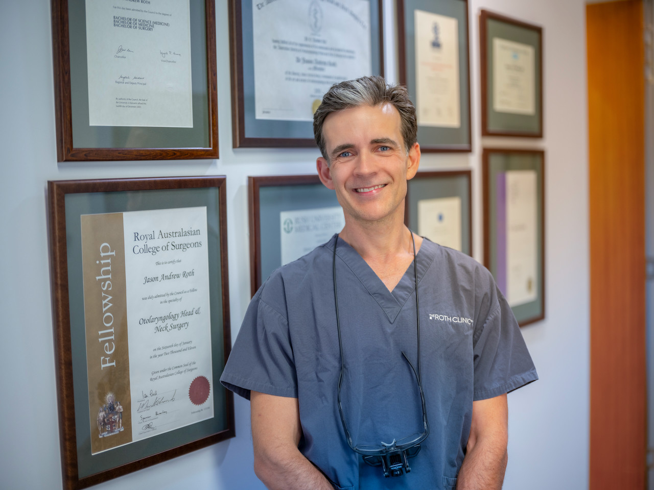
About Dr Jason Roth, MBBS, FRACS

Dr Jason Roth is a Sydney-based Specialist Otolaryngologist & Head and Neck Surgeon with Facial Plastic Surgery Fellowship training from Australia, the United States and Europe. He trained in rhinoplasty under Professor Nolst Trenité in Amsterdam and spent time observing Professor Dean Toriumi in Chicago — two of the most influential rhinoplasty surgeons of the contemporary era.
He attended the inaugural Preservation Rhinoplasty Conference in Nice (2019) and the Structure and Preservation Rhinoplasty Conference in Istanbul (2024). He participates in annual cadaveric rhinoplasty dissection courses through the AAFPS Masters Symposia and uses QMP and equivalent surgical video platforms as part of his ongoing continuing education.
He performs more than 150 rhinoplasty procedures per year and more than 300 nasal surgery procedures per year. View his full profile here →
Rhinoplasty Before & After Gallery
All surgery performed by Dr Jason Roth (MED0001185485), Specialist Otolaryngologist & Head and Neck Surgeon, Sydney. Results vary from person to person. The outcomes shown are relevant only to the specific patient depicted and do not necessarily reflect the results other patients may experience.
View Full Patient Photo Gallery →
If you would like to arrange a consultation, please contact the practice. Two consultations are always required before rhinoplasty proceeds — questions are welcome at every stage of the process.
Preservation Rhinoplasty
- Preservation Rhinoplasty — Overview
- Dorsal Preservation Rhinoplasty
- High Dorsal Strip Rhinoplasty
- Intermediate Strip Rhinoplasty
- Low Dorsal Strip Rhinoplasty
- SPQR Rhinoplasty
- Finocchi SPQR Rhinoplasty
- Preservation vs Open Structure
Structure & Approach
- Open Structure Rhinoplasty
- Closed Approach Rhinoplasty
- Revision Rhinoplasty
- Functional Rhinoplasty
- Non-European Rhinoplasty
- Ultrasonic Rhinoplasty — Disadvantages
Cartilage Grafting
- Rib Grafts
- Allograft vs Own Rib
- Spreader Grafts
- Alar Batten Grafts
- Lateral Crural Strut Grafts
- Nasoseptal Reconstruction
Specific Concerns
- Dorsal Hump
- Wide Nasal Bridge
- Wide Nasal Tip
- Bent or Crooked Nose
- Over-Projected Nose
- Under-Projected Nose
- Pinched Tip
- Hanging Columella
- Uneven Nostrils
- Short Nose
- Narrow Nose
- Nasal Valve Collapse
- Broken Nose
- Saddle Nose Deformity
- Inverted V Deformity
- Pollybeak Deformity
Planning & Patient Information
- Am I a Candidate?
- Pre-Operative Information
- Post-Operative Care
- Rhinoplasty Anaesthesia
- Rhinoplasty Cost
- Insurance Coverage
- Rhinoplasty FAQs
- Planning a Rhinoplasty
- Rhinoplasty Risks
- Choosing a Surgeon
- Recovery — Month by Month
- Rhinoplasty for Men
- Ethnic Rhinoplasty
- Septorhinoplasty
- Before & After Gallery
Specialist Otolaryngologist & Head and Neck Surgeon
Specialist registration — Otorhinolaryngology, Head & Neck Surgery
View full profile
Dr Roth’s Clinical Perspective
Rhinoplasty has been the procedure I have invested the most time in over my career — in formal training, in continuing education, and in daily practice performing over 150 rhinoplasties a year. It is also the procedure that has changed the most. The shift toward preservation rhinoplasty — working within the native dorsal anatomy rather than excising and reconstructing it — has genuinely improved what is achievable and how patients recover, and I have been incorporating these techniques since attending the inaugural preservation course in Nice in 2019.
The most important thing I can say about rhinoplasty consultation is that the surgeon needs to be able to articulate specifically — before the operation — what they are going to do to each anatomical structure and why. A consultation that ends with vague reassurance and a computer image is not a surgical plan. The plan is the set of specific manoeuvres that will be applied to the specific anatomy, and that is what should be discussed and agreed before any commitment is made.
— Dr Jason Roth, MBBS, FRACS (ORL-HNS), IBCFPRS
